
Home
Digestive
Endocrine
Excretory
Integumentary
Lymphatic
Muscular
Nervous
Reproductive
Respiratory
Skeletal
This Week
Useful Mnemonics
Related Links
Science Careers
Pictures
Credits
The
Systems
Notes and Links
The Skeletal SystemDiagrams & Quiz |
Skeletal
I.  Two types of bone
    A. Compact
        1. Forms diaphysis and thinner surfaces of other bones
        2. Haversian Canals- rings in bones that contain blood vessels
        3. Osteocytes receive nutrients/excrete wastes through haversian
           canals via periosteum and endosteum
    B. Cancellous bone
        1. located in epiphysis
        2. contains trabeculae-acts as structural support,  structure
           conserves weight without sacrificing integrity of strength
             a. no blood vessels
             b. have lamellae and osteocytes
II. Ossification-(bone formation)
    A. Two types
        1. Intramembranous ossification
             a. bones of skull
             b. osteoblast begins to produce bone in connective tissue,
                in areas called ossification centers
             c. osteoblasts orientate themselves on connective tissue and
                begin to form trabeculae
             d. trabeculae radiate out and will constantly remodel themselves
                until growth stops or they are replaced with compact bone

        2. Endochondral-(most of skeleton)
             a. forms from cartilage; cartilage cells increase, enlarge, and
                die, then calcify; happens in center of future bone
             b. the outside is vascularized to aid in production; this act
                causes some connective cells to become osteoblasts; they then
                begin a wrapping process that forms the diaphysis
             c. the center of bone undergoes same process; old calcified
                cartilage is removed by osteoclasts
             d. osteoblasts then begin to form new lamellae
             e. osteoclasts clear center to allow bone marrow to form
I. Bone is connective tissue
    A. Determined by extracellular matrix-collagen (ropelike protein),
       proteoglycan (x-mass tree), and other organic molecules.
    B. Contains Calcium and Phosphate
    C. Fromed in thin sheets called lamellae, bone cells called osteocytes
       (between lamella)
    D. Lacunae-holes containing osteocytes
    E. Canaliculi-tiny canals linking osteocytes
II. Types of bone
    A. Long- longer than wide-limbs
    B. short- broad as long-wrist/ankle
    C. Flat-thin, flat- shape skull, ribs, scaupulae
    D. Irregular- facial/vertebrae
III. Types of Marrow
    A. Yellow-fat
    B. Red- synthesis red blood cells
    C. as age increases so does yellow (why?)
IV. Cartilage
    Contains a different amount of collagen/proteoglycans which give
    it the ability to stretch and spirng back.
V. General Terms
    A. Foramen-hole
    B. Canal/meatus tunnel
    C. Fossa-depression
    D. Tuberosity-lump
    E. Process-projection (arch, ridge)
    F. Condyle-where articulation might fit in a joint.
VI. Two types of Bone
    A. Compact
       1. Forms (canellous.)
    B. Cancellous
Bone growth
2 types:
1. Appositional-osteoblasts from between periostem and existing bone.
   Bone increse diamete
2. Endochondral
    A. Occurs in epiphyseal plate (growth plate) grow from middle out.
    B. Left over cartilage is removed by osteoclasts, osteoblasts then
       move in.
    C. Osteoblasts start depositing bone on the lamella
Bone Remodeling
1. Medullary caving increases. Osteoclasts aid in process of moving old
   dead cells out.
Bone Repeair
1. Much like tissue repair. Most cells cause somethign wound. Fibrin aids
   in pulling tissues inside together, thus pulling the bone together.
2. Zone of tissue repair called callus. Osteoblasts form cancellous bone
   to aids in   healing but will become specialized later.
3. Often stronger after a break.
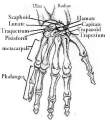
Back to Top Helpful diagrams to learn: 

CLick here to take the Skeletal Quiz. |